$18
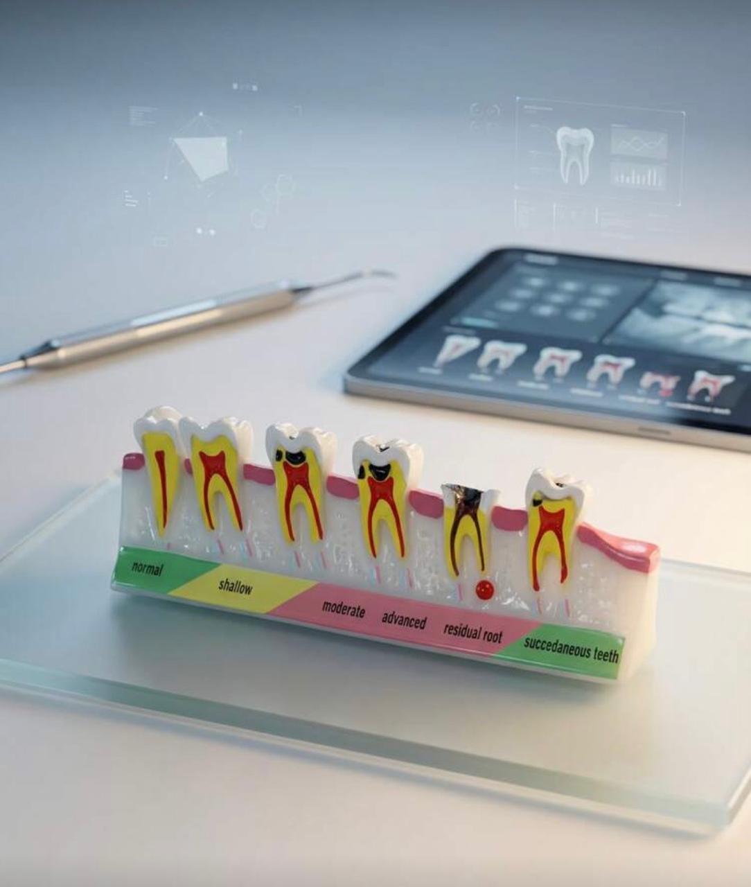
This high-quality dental model clearly illustrates the progressive stages of tooth decay, making it an essential teaching tool for dental professionals, educators, and students. The model visually demonstrates conditions ranging from normal tooth anatomy to shallow decay, moderate decay, advanced decay, residual root, and succedaneous teeth development.
Crafted with detailed cross-sectional views, each tooth highlights internal structures including enamel, dentin, and pulp to enhance patient education and classroom learning. The color-coded base provides clear stage identification, helping users easily explain diagnosis and treatment planning.
Key Features:
Detailed cross-sectional tooth anatomy
Displays multiple stages of dental caries progression
Color-coded labeling for easy understanding
Durable, high-quality construction
Ideal for dental clinics, universities, and training centers
Perfect for chairside patient education or academic demonstrations, this model helps simplify complex dental concepts and improves communication and understanding.